Deprecated: Using ${var} in strings is deprecated, use {$var} instead in /home/u541663480/domains/shanthinethralaya.com/public_html/wp-content/plugins/depicter/vendor/averta/wpemerge-app-core/src/Assets/Assets.php on line 184
Dr.B.Shanthi Niketh | SHANTHINETHRALAYA EYE HOSPITAL

SHANTHINETHRALAYA EYE HOSPITAL

Welcome to Shanthi Nethralaya Eye Hospital – Hyderabad’s Best EyeHospital

Shanthi Nethralaya Eye Hospital is Hyderabad’s best eye hospital, committed to delivering quality, compassionate, and accessible eye care. Led by Dr. B. Shanthi Niketh, recognized as one of Uppal and Hyderabad’s best eye doctors, our hospital provides a full range of services, from routine eye check-ups to advanced eye surgeries. Discover why patients choose Shanthi Nethralaya Eye Hospital as the best choice for eye care in Hyderabad, where affordability meets top-notch care every day.

Renowned for providing the best cataract surgery in Hyderabad, Shanthi Nethralaya Eye Hospital also specializes in vision correction surgeries and spectacle removal surgery . Our advanced cataract procedures restore clear vision with precision, while our spectacle removal options offer freedom from glasses. Choose Shanthi Nethralaya Eye Hospital as your premier destination for advanced cataract surgery and vision correction (spectacle removal) surgery. Under the expertise of Dr. B. Shanthi Niketh, Uppal, Hyderabad’s top eye surgeons, we are dedicated to safe, effective, and personalized care, making us the preferred choice for Advanced Cataract Surgeries and vision correction (Spectacle/glasses removal) surgeries in Hyderabad.

Your Vision Is Our Top Priority—Experience the Best Eye Care, Because We Treat Your Eyes Like Our Own!"

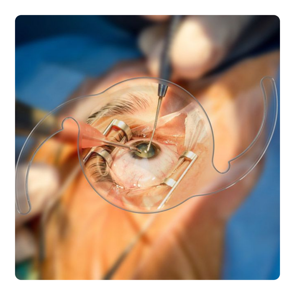
"Surgeon performing cataract surgery using a microscope."

Welcome to Shanthi Nethralaya Eye Hospital, where we’re proud to be one of the best eye hospitals in Hyderabad. Our focus is on you, our valued patient, as we deliver personalized, safe, and high-quality care tailored to your unique needs.Our state-of-the-art facility spans a spacious 6,000-square-foot building and boasts the latest technology, including a modular operation theatre, to provide you with the very best eye treatments available.Our commitment goes far beyond our advanced facilities. We create a welcoming, comfortable environment where each patient receives the dedicated attention and care they deserve. Whether you’re here for a routine eye examination or a complex surgical procedure, our experienced professionals are by your side every step.At Shanthi Nethralaya Eye Hospital, we strive to be the best hospital for your eye care needs. We empower you with knowledge and options so you feel confident in your care and treatment plans. Your well-being and satisfaction are our top priorities.Come experience the difference of compassionate, world-class eye care with us. Let us guide you on your journey to a better vision and improved quality of life.

Welcome to Shanthi Nethralaya Eye Hospital, where we’re proud to be one of the best eye hospitals in Hyderabad. Our focus is on you, our valued patient, as we deliver personalized, safe, and high-quality care tailored to your unique needs.Our state-of-the-art facility spans a spacious 6,000-square-foot building and boasts the latest technology, including a modular operation theatre, to provide you with the very best eye treatments available.

A Leading Expert In Cornea & Refractive Surgery

MBBS,DO,FICO,FLVPEI

Consultant Ophthalmologist

Cataract,Cornea & Refractive Surgeon

What patients Say About Us

Why Choose Shanthi Nethralaya Eye Hospital?

Best Patient Outcome

We strive to achieve the best possible outcomes for each patient through our tailored treatment plans and dedicated care. Our focus on excellence ensures you receive effective, life-changing results.

Modular Operation Theatre

Our hospital is equipped with a state-of-the-art modular operation theatre, providing an optimal environment for safe and efficient surgeries. This advanced setup allows us to deliver the best possible outcomes for our patients.

Affordable Eye Care

We understand the importance of providing quality eye care that is accessible to everyone. Our services are offered at affordable prices, ensuring that patients from all walks of life can receive the care they need without financial stress.

One Patient at a Time

At Shanthi Nethralaya Eye Hospital, we believe in giving our undivided attention to each patient during examinations. This approach ensures that you receive personalized care and detailed attention.

Patient Confidentiality

We take your privacy seriously and uphold the highest standards of patient confidentiality. You can trust us to handle your medical information with care and respect at all times.

Advanced Facilities and Equipment

Shanthi Nethralaya Eye Hospital is equipped with the latest diagnostic and treatment technologies, ensuring precise and efficient eye care. Our advanced imaging and surgical tools support the highest standard of ophthalmic care.

Why Choose Shanthi Nethralaya Eye Hospital ?

OUR CORE VALUES

F.A.Q.

Choose Shanthi Nethralaya Eye Hospital for unparalleled eye care excellence. Conveniently located and easily accessible, our hospital ensures that patients from all walks of life can receive top-notch services without financial strain. With a state-of-the-art modular operation theatre, led by Dr. B. Shanthi Niketh, we deliver safe and efficient surgeries, focusing on achieving the best outcomes for every patient. Our personalized approach guarantees individualized attention and confidentiality, ensuring your comfort and trust. Dr. B. Shanthi Niketh, a highly skilled ophthalmologist, leads our team in offering advanced treatments and solutions for all eye conditions, including cataract surgeries with the best outcomes and vision correction surgeries. From the warm welcome at our doors to the comprehensive range of services provided under one roof, Shanthi Nethralaya Eye Hospital sets the standard for excellence in eye care. Choose us for a transformative experience and the best patient outcomes.

1)Open the Google Maps app on your smartphone or visit the Google Maps website on your computer browser.

2)In the search bar, type “Shanthi Nethralaya Eye Hospital” and press Enter or tap on the search icon.

3)Google Maps will display the location of Shanthi Nethralaya Eye Hospital on the map, along with additional information such as address, contact details, and reviews.

4)If you’re using the Google Maps app on your smartphone, you can tap the “Directions” button to get directions to the hospital from your current location.

1)While traveling on NH163, also known as Hyderabad Warangal Highway, towards Warangal.

2)After about 1.2 km from Uppal ‘x’ road, make a U-turn just before the Asian Multiplex Theatre.

3)Take a left turn near Darbar Bar and Restaurant. You’ll also notice it nearby.

4)After taking the left turn at Darbar Bar and Restaurant, proceed ahead and take the first right.

5)You will then see Shanthi Nethralaya Eye Hospital on the left-hand side.

These simplified directions will guide you to Shanthi Nethralaya Eye Hospital effectively.

Choose Shanthi Nethralaya Eye Hospital for your cataract surgery, where you’ll experience unparalleled expertise and cutting-edge facilities. Under the guidance of Dr. B. Shanthi Niketh, a distinguished expert in cornea and refractive surgery, we offer personalized care and surgical precision. Our modular operation theatre, featuring HEPA filter unidirectional laminar airflow, ensures the highest safety standards. With state-of-the-art equipment and Dr. Shanthi Niketh’s leadership, we deliver optimal outcomes for each patient. Our unwavering commitment to excellence and patient-centric care guarantees a transformative journey, leading to improved vision and enhanced quality of life. Trust Shanthi Nethralaya Eye Hospital for your cataract surgery, and embark on a path to clearer vision and better eye health.

Choose Dr. B. Shanthi Niketh as your preferred surgeon for refractive or LASIK surgery for unparalleled expertise and exceptional outcomes. With a reputation as the best eye doctor and cataract surgeon, Dr. Shanthi Niketh specializes in cornea and refractive surgery, backed by extensive training and over 5000 successful cataract surgeries. As Head of the Cornea Department at Akira Eye Hospital and Chief Cornea Consultant at Neoretina Eye Hospital, he ensures the highest standards of clinical excellence and patient-centric care. Dr. Shanthi Niketh’s personalized treatment plans and commitment to optimal outcomes make him the ideal choice for your vision correction needs. Trust in his expertise and experience to deliver transformative results and enhanced visual clarity.

At Shanthi Nethralaya Eye Hospital, the consultation fee is 300 rupees, which covers a comprehensive eye examination. This includes a doctor consultation, vision testing/refraction, slit-lamp examination, IOP measurement, glaucoma screening, and diabetic retinopathy screening.

Welcome to Shanthi Nethralaya Eye Hospital, where we prioritize your eye health and well-being. Led by Dr. B. Shanthi Niketh, a renowned expert in cornea and refractive surgery, our hospital offers personalized, safe, and high-quality care tailored to your unique needs. With a spacious 6,000-square-foot facility equipped with the latest technology, including a modular operation theatre, we ensure optimal treatment outcomes. Our commitment to excellence extends to our welcoming environment, where each patient receives dedicated attention and care. At Shanthi Nethralaya Eye Hospital, we strive for the best patient outcomes through tailored treatment plans and expert care from our highly skilled medical team. With advanced facilities and comprehensive services, we provide all your eye care needs under one roof. Choose us as your healthcare partner for a transformative experience in eye care.

Ready to See Clearly? Take the First Step Towards a Better Vision Today!

Call
+91-8074661750

Author name: Dr.B.Shanthi Niketh

A man experiencing eye strain, holding his glasses in one hand and rubbing his eyes, seated in front of a laptop.

Computer Vision Syndrome

Menu Home About Us Doctor Vision & Mission Services Advanced Cataract Services Cornea Vision Correction or Spectacles Removal Surgery Glaucoma Pediatric Ophthalmology Occuloplasty Medical Retina Opticals Conditions We Treat Allergic conjunctivitis Keratoconus Dry Eyes Patient Education Blog Posters Videos Gallery Computer Vision Syndrome In today’s digital age, many people spend a significant portion of their day in front of screens, whether it’s a computer, tablet, or smartphone. This extended screen time can lead to a range of vision-related problems collectively known as Computer Vision Syndrome (CVS), also referred to as digital eye strain. Understanding CVS is crucial for maintaining healthy vision and preventing discomfort while working or using digital devices. What is Computer Vision Syndrome? Computer Vision Syndrome is a collection of eye and vision-related problems that result from prolonged use of digital screens. Symptoms can arise after just a few hours of screen exposure and may worsen with continued use. CVS affects individuals of all ages, but it is particularly common among those who work long hours at a computer. Common Symptoms of Computer Vision Syndrome Eye Strain: A feeling of discomfort or fatigue in the eyes. Dry Eyes: Reduced blinking while staring at screens can lead to dryness and irritation. Blurred Vision: Difficulty focusing on the screen, especially after prolonged use. Headaches: Tension headaches can develop as a result of eye strain and poor posture. Neck and Shoulder Pain: Poor ergonomics while using a computer can lead to discomfort in the neck and shoulders. Difficulty Focusing: Trouble switching focus between the screen and other objects. Causes of Computer Vision Syndrome Extended Screen Time: Spending long hours staring at digital devices without breaks. Poor Lighting: Glare from overhead lighting or reflections on the screen can strain the eyes. Improper Screen Position: Screens that are too high, low, or at awkward angles can lead to poor posture and discomfort. Inadequate Blink Rate: When focused on screens, people tend to blink less, leading to dryness and irritation. Uncorrected Vision Problems: Pre-existing vision issues like nearsightedness, farsightedness, or astigmatism can exacerbate symptoms. Prevention and Treatment Strategies Follow the 20-20-20 Rule Every 20 minutes, take a break and look at something 20 feet away for at least 20 seconds. This helps relax the eye muscles and reduces strain. Adjust Your Workstation Screen Position: Position the screen at eye level and about an arm’s length away to maintain a comfortable viewing angle. Chair and Desk Height: Ensure your chair supports your lower back and your desk is at a height that allows your feet to rest flat on the floor. Use Proper Lighting Reduce glare by using screens with anti-reflective coatings and adjusting room lighting. Natural light is preferable, so position your screen away from direct sunlight. Blink Regularly Consciously blinking can help keep the eyes moist and reduce dryness. Consider using artificial tears if necessary. Get Regular Eye Exams Visit an eye care professional to ensure your vision is corrected adequately. They can also recommend special lenses designed to reduce digital eye strain. When to Seek Professional Help If symptoms persist despite taking preventive measures, it may be time to consult an eye care professional. They can conduct a comprehensive eye examination and provide tailored solutions to address your specific needs, such as prescription glasses for computer use or advice on managing screen time. Conclusion Computer Vision Syndrome is a common issue in our screen-dominated world, affecting many individuals daily. By understanding its causes and symptoms, as well as adopting preventative measures, you can protect your vision and enhance your comfort while using digital devices. Why Choose Dr. Shanthi Niketh and Shanthi Nethralaya for Computer & Digital Eye Strain Issues Expertise in Ophthalmology Dr. Shanthi Niketh is a highly trained ophthalmologist with extensive experience in diagnosing and treating a wide range of eye conditions, including computer vision syndrome. His knowledge of the latest advancements in eye care ensures that patients receive the best treatment options available. Comprehensive Eye Examinations At Shanthi Nethralaya, we provide thorough eye examinations that go beyond just checking vision. We evaluate eye health in detail, assessing factors contributing to digital eye strain, such as tear film quality, eye muscle coordination, and overall ocular health. Personalized Treatment Plans Every patient is unique, and so are their eye care needs. Dr. Shanthi Niketh creates customized treatment plans tailored to each individual’s specific symptoms and lifestyle. Whether it’s prescribing the right glasses, recommending digital eye exercises, or suggesting lifestyle modifications, our approach is patient-centered. State-of-the-Art Facilities Shanthi Nethralaya is equipped with modern diagnostic tools and technology to accurately assess and manage computer vision syndrome. Our advanced equipment allows for precise measurements and evaluations to ensure effective treatment. Personalized Treatment Plans Every patient is unique, and so are their eye care needs. Dr. Shanthi Niketh creates customized treatment plans tailored to each individual’s specific symptoms and lifestyle. Whether it’s prescribing the right glasses, recommending digital eye exercises, or suggesting lifestyle modifications, our approach is patient-centered. State-of-the-Art Facilities Shanthi Nethralaya is equipped with modern diagnostic tools and technology to accurately assess and manage computer vision syndrome. Our advanced equipment allows for precise measurements and evaluations to ensure effective treatment. Focus on Preventive Care We emphasize the importance of preventive measures to avoid digital eye strain. Dr. Shanthi Niketh educates patients on proper ergonomics, the significance of regular breaks, and strategies to optimize screen use, empowering them to take control of their eye health. Patient-Centric Environment Our hospital prioritizes patient comfort and satisfaction. The staff is trained to provide a welcoming atmosphere, ensuring that each visit is pleasant and stress-free. Dr. Shanthi Niketh and the team are always available to address patient concerns and provide guidance throughout the treatment process. Ongoing Support and Follow-Up After treatment, we offer continued support and follow-up appointments to monitor progress and make necessary adjustments to treatment plans. Our commitment to our patients extends beyond the initial visit, ensuring they achieve long-term eye health. Community Engagement Shanthi Nethralaya actively participates in community awareness programs about eye health, particularly concerning

Close-up of an elderly person's eye, highlighting wrinkles, fine lines, and signs of aging around the eye.

Geriatric Eye Diseases

Menu Home About Us Doctor Vision & Mission Services Advanced Cataract Services Cornea Vision Correction or Spectacles Removal Surgery Glaucoma Pediatric Ophthalmology Occuloplasty Medical Retina Opticals Conditions We Treat Allergic conjunctivitis Keratoconus Dry Eyes Patient Education Blog Posters Videos Gallery Common Geriatric Eye Diseases Cataracts What Are They?: A cataract is a clouding of the eye’s natural lens, leading to blurred vision. Symptoms: Patients may experience difficulty seeing at night, sensitivity to light, and faded colors. Treatment: Surgery is the most effective treatment, where the cloudy lens is replaced with an artificial lens. Age-Related Macular Degeneration (AMD) What Is It?: AMD affects the macula, the part of the retina responsible for sharp vision. Symptoms: Early signs include blurred vision and difficulty recognizing faces; advanced AMD can lead to significant vision loss. Treatment: While there’s no cure, treatments like injections and laser therapy can help manage the condition. Glaucoma What Is It?: Glaucoma refers to a group of diseases that damage the optic nerve, often due to increased eye pressure. Symptoms: Many people experience no symptoms until significant vision loss occurs, but peripheral vision loss and halos around lights can occur. Treatment: Options include prescription eye drops, oral medications, and surgical procedures to lower eye pressure. Diabetic Retinopathy What Is It?: This disease affects the blood vessels in the retina of those with diabetes. Symptoms: Early stages may be symptomless; as it progresses, symptoms like blurred vision and floaters may develop. Treatment: Managing diabetes and treatments like laser surgery can help prevent vision loss. Dry Eye Syndrome What Is It?: Aging can lead to reduced tear production, causing dry, irritated eyes. Symptoms: Patients often report dryness, burning sensations, and redness. Treatment: Artificial tears and lifestyle modifications can alleviate symptoms. Retinal Detachment Call +91-8074661750 What Is It?: This serious condition occurs when the retina separates from the back of the eye. Symptoms: Sudden flashes of light, floaters, and a shadow over vision may indicate this condition. Treatment: Immediate medical attention is essential; treatment may involve laser surgery or cryotherapy. Tips for Maintaining Eye Health in Old Age Regular Eye Exams: Schedule annual check-ups with an eye care professional to detect issues early. Healthy Diet: Consume foods rich in antioxidants and omega-3 fatty acids, such as leafy greens and fish. Protect Your Eyes: Wear UV-protected sunglasses outdoors to shield your eyes from harmful rays. Manage Chronic Conditions: Keep diabetes and hypertension under control to protect eye health. Quit Smoking: Smoking cessation reduces the risk of cataracts and AMD. Limit Screen Time: Follow the 20-20-20 rule to reduce digital eye strain. Why Choose Shanthi Nethralaya? Expert Care: Led by Dr. Shanthi Niketh, our experienced team provides specialized care for geriatric eye diseases. Comprehensive Eye Exams: We conduct thorough eye examinations to detect and treat age-related eye problems. Personalized Treatment Plans: Every patient receives a tailored approach based on their specific needs and conditions. State-of-the-Art Facilities: Our advanced technology allows for accurate diagnosis and effective treatment options. Patient-Centric Approach: We prioritize patient comfort and satisfaction, providing a welcoming environment and ongoing support. Conclusion Understanding and addressing geriatric eye diseases is vital for maintaining vision and quality of life in older adults. At Shanthi Nethralaya, we are committed to providing comprehensive eye care tailored to the unique needs of our patients. If you or a loved one is experiencing vision changes, contact us today for an eye examination. Whatsapp Icon-email Instagram Youtube Icon-map-marker1 Services & Treatments Cataract Cornea Glaucoma Vision Correction Pediatric Ophthalmology Occuloplasty Medical Retina Opticals Contact info Hanumasai Nagar, Shanti Nagar, Uppal, Hyderabad, Telangana 500039, India +91 80746 61750 shanthinethralaya@gmail.com Opening Hours: 09:00 AM – 08:00 PM Social Profiles Instagram Whatsapp Youtube Icon-map-marker

Close-up of an eye showing signs of herpes zoster ophthalmicus (HZO), with redness, inflammation, and corneal scarring visible.

Herpes Zoster ophthalmicus(HZO)

Menu Home About Us Doctor Vision & Mission Services Advanced Cataract Services Cornea Glaucoma Vision Correction/Refractive Surgery Pediatric Ophthalmology Occuloplasty Medical Retina Opticals Conditions We Treat Allergic conjunctivitis Keratoconus Dry Eyes Patient Education Blog Posters Videos Gallery Herpes Zoster ophthalmicus Herpes Zoster Ophthalmicus (HZO), also known as shingles in the eye, is a viral infection caused by the reactivation of the varicella-zoster virus—the same virus responsible for chickenpox. When the virus reactivates, it can affect the skin around the eye and, in some cases, the eye itself. This condition can cause significant discomfort and, if not treated, lead to complications like vision loss. What Causes Herpes Zoster Ophthalmicus? HZO is caused by the varicella-zoster virus, which lies dormant in the nerve cells after someone has recovered from chickenpox. Years or even decades later, the virus can reactivate due to a weakened immune system, stress, or aging, causing shingles. When the ophthalmic division of the trigeminal nerve (the nerve that controls sensation in the face) is affected, it leads to Herpes Zoster Ophthalmicus. Who is More Prone to Herpes Zoster Ophthalmicus? While anyone who has had chickenpox can develop HZO, certain individuals are at a higher risk of experiencing this viral reactivation. Here are some of the factors that increase susceptibility Older Adults: People over the age of 50 are at a higher risk, as the immune system weakens with age, making it easier for the virus to reactivate. Weakened Immune System: Conditions that compromise the immune system, such as HIV/AIDS, cancer, or autoimmune diseases, can make individuals more prone to HZO. Those undergoing chemotherapy or radiation therapy are also at increased risk. Use of Immunosuppressive Medications: Individuals on medications like steroids or drugs used after organ transplants (e.g., to prevent organ rejection) have an increased risk of developing HZO. Stress and Fatigue: Physical and emotional stress, combined with prolonged fatigue, can weaken the immune system, triggering a reactivation of the virus. History of Chickenpox or Shingles: If you’ve previously had chickenpox or shingles, you are at risk of developing Herpes Zoster Ophthalmicus as the virus remains dormant in the body. Symptoms of HZO Pain and Tingling: The initial symptom is often a tingling, burning sensation, or pain on one side of the forehead or around the eye. Rash: A red rash or small blisters typically appear on the forehead, eyelids, and sometimes the tip of the nose. Eye Involvement: Redness, swelling, light sensitivity, and eye pain may develop if the virus affects the eye itself. Blurred Vision: Vision may become blurry or hazy, especially if the cornea (the clear front part of the eye) is involved. Fever and Fatigue: Some people may experience general symptoms like fever and feeling tired. How is Herpes Zoster Ophthalmicus Diagnosed? HZO is usually diagnosed based on clinical signs and symptoms. An ophthalmologist will look for the characteristic rash and may perform an eye examination to check for any involvement of the cornea, retina, or optic nerve. Slit-lamp examination: To look closely at the structures inside the eye. Visual acuity test: To check for any changes in vision. Blood tests or viral cultures: In some cases, these may be used to confirm the presence of the varicella-zoster virus. Complications of Herpes Zoster Ophthalmicus Corneal Scarring: This can lead to permanent vision loss if the cornea is damaged. Glaucoma: Increased pressure in the eye can occur due to inflammation. Postherpetic Neuralgia: Even after the rash heals, some individuals may experience long-term nerve pain in the affected area. Vision Loss: Severe cases can lead to partial or total vision loss if the eye is significantly damaged. Treatment Options for Herpes Zoster Ophthalmicus Antiviral Medications Acyclovir, Valacyclovir, or Famciclovir are commonly prescribed antiviral drugs that help shorten the duration of the infection and lessen its severity. These medications are most effective when started within 72 hours of the onset of symptoms. Corticosteroids Eye drops or oral corticosteroids may be prescribed to reduce inflammation in the eye. However, they should only be used under the guidance of an eye doctor. Pain Relievers Over-the-counter pain medications or prescription painkillers may be used to alleviate discomfort during the active phase of the infection. Lubricating Eye Drops: Artificial tears can help with dryness and discomfort caused by the inflammation of the eye. Treatment for Postherpetic Neuralgia If long-term nerve pain persists after the rash heals, your doctor may prescribe pain medications, nerve blocks, or other therapies to manage the pain. Prevention of Herpes Zoster Ophthalmicus One of the most effective ways to prevent HZO is through vaccination. The shingles vaccine (Zoster vaccine) is recommended for adults over 50, as it can significantly reduce the risk of developing shingles and HZO. If you’ve had chickenpox or shingles before, getting vaccinated can still lower your chances of a recurrence. Why Choose Shanthi Nethralaya for Herpes Zoster Ophthalmicus Treatment? At Shanthi Nethralaya, we offer personalized care for patients with Herpes Zoster Ophthalmicus. Our well-trained optometrists and ophthalmologists provide thorough eye examinations, ensuring early detection and treatment of this condition. With a commitment to using the latest technology and treatment options, our experienced team helps manage the infection and prevent long-term complications like vision loss. One Patient at a Time : At Shanthi Nethralaya, every patient is given individual attention during their consultation, ensuring that all your concerns are addressed. Whatsapp Icon-email Instagram Youtube Icon-map-marker1 Call +91-8074661750 Services & Treatments Cataract Cornea Glaucoma Vision Correction Pediatric Ophthalmology Occuloplasty Medical Retina Opticals Contact info Hanumasai Nagar, Shanti Nagar, Uppal, Hyderabad, Telangana 500039, India +91 80746 61750 shanthinethralaya@gmail.com Opening Hours: 09:00 AM – 08:00 PM Social Profiles Instagram Whatsapp Youtube Icon-map-marker

"Diagram of the human eye showing the focus of light on the retina, representing refractive errors like myopia, hyperopia, and astigmatism."

Refractive Errors:Myopia, Hyperopia and Astigmatism

Menu Home About Us Doctor Vision & Mission Services Advanced Cataract Services Cornea Glaucoma Vision Correction/Refractive Surgery Pediatric Ophthalmology Occuloplasty Medical Retina Opticals Conditions We Treat Allergic conjunctivitis Keratoconus Dry Eyes Patient Education Blog Posters Videos Gallery Understanding Refractive Errors: Myopia, Hyperopia & Astigmatism Having clear vision depends on how well your eyes can focus light onto the retina. Refractive errors occur when the shape of your eye prevents light from focusing correctly. This results in blurry vision, which can be easily corrected with glasses, contact lenses, or surgery. Let’s take a look at the four most common refractive errors: myopia, hyperopia and astigmatism. Whatsapp Icon-email Instagram Youtube Icon-map-marker1 What is Myopia (Nearsightedness)? Myopia, commonly known as nearsightedness, occurs when you can see objects close to you clearly but struggle to focus on distant objects. This happens because light entering your eye focuses in front of the retina instead of directly on it. Signs of Myopia Call +91-8074661750 Difficulty seeing distant objects clearly (e.g., the blackboard or road signs). Squinting to see better. Frequent headaches due to eye strain. Correction for Myopia Myopia is corrected using minus power lenses (concave lenses). These lenses help focus light directly on the retina, improving distance vision. What is Hyperopia (Farsightedness)? Hyperopia, or farsightedness, is the opposite of myopia. People with hyperopia can see distant objects clearly but may struggle with close-up tasks like reading. In this condition, light focuses behind the retina. Signs of Hyperopia Difficulty focusing on nearby objects, such as books or screens. Eye strain, especially when reading or doing detailed work. Blurry vision when looking at objects up close. Correction for Hyperopia Hyperopia is corrected using plus power lenses (convex lenses). These lenses adjust the focus so that light lands on the retina, allowing for clear near vision. What is Astigmatism? Astigmatism is corrected with cylindrical lenses, which have different powers in different areas to correct the uneven curvature of the eye. Why Should You Get Your Eyes Checked for Refractive Errors? If you experience any signs of blurry vision, eye strain, or difficulty seeing objects clearly, it’s important to get a comprehensive eye examination. Regular eye check-ups ensure that any vision problems are detected early and corrected effectively. Why Choose Shanthi Nethralaya for Your Routine Eye Examination? At Shanthi Nethralaya, we believe in providing personalized care, one patient at a time. Every patient undergoes a thorough examination by both a highly trained optometrist (technician) and an ophthalmologist (eye doctor). Our team ensures that no detail is missed in your diagnosis and treatment. We offer comprehensive eye care services with a focus on precise diagnosis and personalized treatment for refractive errors. Our experienced team uses the latest technology to ensure accurate prescriptions for glasses or contact lenses. Whether you need a routine eye exam or specialized care, we are here to provide the highest standard of eye care. Services & Treatments Cataract Cornea Glaucoma Vision Correction Pediatric Ophthalmology Occuloplasty Medical Retina Opticals Contact info Hanumasai Nagar, Shanti Nagar, Uppal, Hyderabad, Telangana 500039, India +91 80746 61750 shanthinethralaya@gmail.com Opening Hours: 09:00 AM – 08:00 PM Social Profiles Instagram Whatsapp Youtube Icon-map-marker

Child with eye injury due to Diwali firecracker accident

Keep Your Eyes Safe This Diwali : Firecracker Safety Tips

Menu Home About Us Doctor Vision & Mission Services Advanced Cataract Services Cornea Glaucoma Vision Correction/Refractive Surgery Pediatric Ophthalmology Occuloplasty Medical Retina Opticals Conditions We Treat Allergic conjunctivitis Keratoconus Dry Eyes Patient Education Blog Posters Videos Gallery Keep Your Eyes Safe This Diwali : Firecracker Safety Tips Firecrackers are an integral part of Diwali celebrations, but they come with risks, especially to the eyes. Each year, thousands of people sustain eye injuries due to fireworks. Shanthi Nethralaya Eye Hospital, known for its expertise in handling eye emergencies, strongly advises following safety measures to avoid such mishaps. Here’s a complete guide on how to prepare before starting the celebration, what to do in case of an eye injury, and when to seek medical help. Before Starting the Celebration: Precautions to Take Wear Protective Eyewear Always wear safety goggles or even basic sunglasses to protect your eyes from sparks and debris. Ensure children and others around you are wearing eye protection too, especially if they’re handling fireworks. Keep a Safe Distance Maintain a distance of at least 5-10 feet when lighting fireworks. Avoid standing too close to where others are setting off fireworks. Use a Long-Stemmed Lighter Use a candle or long-stemmed lighter to light firecrackers. This gives you enough distance from the cracker to avoid sudden bursts. Have a First Aid Kit and Water Ready Keep a first aid kit handy, especially with saline solution, sterile eye pads, and water bottles. In case of any eye injury, it’s important to act quickly. Having water ready can help wash away particles or chemicals immediately. Ensure Proper Lighting in the Area Always light firecrackers in a well-lit area to avoid accidents. Poor lighting can make it harder to see the direction of sparks or falling debris. What to Do in Case of an Eye Injury If Debris or Sparks Enter the Eye Flush the Eye with Water First, avoid rubbing your eyes as this can cause further damage. Wash your hands thoroughly if you’ve been handling firecrackers. Flush the eye gently with clean water. Running water from a tap is ideal. Alternative: Use a Water Bottle If running water isn’t available, grab a clean water bottle. Poke a few small holes in the cap, then squeeze the bottle gently to create a stream of water. Wash your eye thoroughly to flush out any foreign particles. Use Saline Solution If you have saline solution available, use it to rinse your eye. Tilt your head to the side and pour the solution across your eye to wash away any debris. In Case of a Blunt Injury to the Eye (Impact from Fireworks) Do not apply pressure on the injured eye. Blunt trauma can cause internal damage that might not be visible at first. Place a cold compress over the eye to reduce swelling and pain. Use a soft, clean cloth and cold water or ice wrapped in cloth. Seek medical attention immediately. You can visit Shanthi Nethralaya Eye Hospital, where Dr. Shanthi Niketh, a leading Cornea Specialist, can provide prompt and expert care for any serious eye injuries. In Case of an Open-Globe Injury (Cut or Tear in the Eye) If there is a cut or tear in the eye from firecrackers, avoid touching or pressing the area. Do not try to remove any embedded particles from the eye. Cover the eye with a clean, sterile eye pad or soft cloth to protect it until you get medical help. See a doctor immediately, as an open-globe injury requires emergency medical treatment to prevent loss of vision. When to See a Doctor Persistent Pain or Discomfort If the pain continues for more than a few minutes after washing your eyes, it could indicate a deeper injury or foreign particle still in the eye. Blurry or Impaired Vision If you notice blurry vision, double vision, or loss of vision after an injury, this may be a sign of serious internal damage. Visible Cuts, Tears, or Bleeding Any visible tear, cut, or bleeding around the eye or eyelid should be treated as an emergency. Apply a sterile dressing and see a doctor immediately. Excessive Swelling or Bruising Swelling or bruising that doesn’t subside or increases in intensity should be evaluated by a doctor to rule out internal damage. Sensitivity to Light If you develop light sensitivity (photophobia) after an injury, this can be a sign of damage to the eye or retina. Immediate medical attention is necessary. Conclusion Celebrating Diwali with firecrackers is a joyous tradition, but safety should always come first, especially when it comes to protecting your eyes. By taking precautions like wearing protective eyewear, keeping a safe distance, and knowing how to respond in case of injury, you can enjoy the festivities without the risk of injury. Remember, for serious eye injuries, it’s always best to consult a doctor as soon as possible to prevent long-term damage. Stay safe, and make sure to visit Shanthi Nethralaya Eye Hospital if you require expert care for any firecracker-related eye injuries.

"Collage of various causes of cataract including aging, diabetes, smoking, eye trauma, medications, and genetic factors, with text overlay 'Causes of Cataract'.

Causes of Cataract and How to Delay Its Progression

Menu Home About Us Doctor Vision & Mission Services Advanced Cataract Services Cornea Glaucoma Vision Correction/Refractive Surgery Pediatric Ophthalmology Occuloplasty Medical Retina Opticals Conditions We Treat Allergic conjunctivitis Keratoconus Dry Eyes Patient Education Blog Posters Videos Gallery Cataracts Cataracts are a common eye condition, especially as we age, leading to cloudy or blurry vision. Although age-related cataracts are inevitable for many, they don’t progress at the same rate for everyone. Just like the way our hair turns white as we age, cataracts form at different stages depending on various factors. In this post, we’ll explore the causes of cataracts and practical tips to delay their progression, particularly focusing on age-related cataracts. What Causes Cataracts? Age The natural aging process is the leading cause, typically affecting people over 60. Genetics A family history of cataracts may increase your likelihood of developing them earlier. Health Conditions Diabetes, hypertension, and other medical conditions can accelerate cataract formation. Sun Exposure Prolonged exposure to UV rays can damage the lens, hastening cataract development. Smoking and Alcohol These lifestyle habits increase oxidative stress, contributing to cataract formation. Eye Injuries or Surgeries Trauma or previous surgeries to the eye can lead to cataracts over time. The Analogy: Aging and Cataracts, Like White Hair Just as aging causes some people to get white hair earlier than others, cataracts also progress at different rates for different people. Some individuals may notice white hair in their 30s, while others won’t experience it until much later in life. Similarly, cataracts can develop earlier in some due to lifestyle, genetics, or health conditions, while others may maintain clear vision into their 60s or 70s. The key is recognizing the risk factors and taking steps to delay the progression, much like how some people try to maintain healthy hair by managing stress, diet, and overall health. How to Delay Cataract Progression Wear UV Protection Long-term exposure to the sun’s harmful UV rays accelerates cataract development. Wear sunglasses with UV protection when outdoors to shield your eyes from damage. Call +91-8074661750 Eat an Antioxidant-Rich Diet A diet rich in antioxidants such as Vitamin C, Vitamin E, and beta-carotene can help slow the progression of cataracts by protecting your eyes from oxidative damage. Incorporate leafy greens, citrus fruits, carrots, and nuts into your daily meals. Quit Smoking and Limit Alcohol Smoking and excessive alcohol consumption increase the production of free radicals, which can harm the lens. Quitting smoking and limiting alcohol intake can reduce your risk of early cataract development Whatsapp Icon-email Instagram Youtube Icon-map-marker1 Manage Health Conditions Chronic conditions like diabetes are known to accelerate cataract formation. Keeping blood sugar and blood pressure under control can help delay cataract progression. Regular Eye Exams Routine eye check-ups are crucial, especially after the age of 40. Early detection can help monitor cataracts and take preventive measures to slow their development. Why Choose Dr. Shanthi Niketh and Shanthi Nethralaya Eye Hospital for Cataract Surgery? At Shanthi Nethralaya Eye Hospital, Dr. Shanthi Niketh is recognized as Hyderabad’s best eye surgeon, specializing in advanced cataract surgery. With years of expertise and a patient-centered approach, Dr. B.Shanthi Niketh ensures that every patient receives personalized care and the latest surgical techniques for optimal vision outcomes. Our state-of-the-art facilities, combined with compassionate care, make Shanthi Nethralaya the best choice for cataract surgery Conclusion: Take Charge of Your Eye Health While aging is an inevitable factor in cataract formation, proactive steps can delay its progression. By protecting your eyes and maintaining overall health, you can enjoy clearer vision for longer. And when the time comes for cataract surgery, trust Dr. Shanthi Niketh and Shanthi Nethralaya Eye Hospital to provide world-class care and exceptional results. Services & Treatments Cataract Cornea Glaucoma Vision Correction Pediatric Ophthalmology Occuloplasty Medical Retina Opticals Contact info Hanumasai Nagar, Shanti Nagar, Uppal, Hyderabad, Telangana 500039, India +91 80746 61750 shanthinethralaya@gmail.com Opening Hours: 09:00 AM – 08:00 PM Social Profiles Instagram Whatsapp Youtube Icon-map-marker

Symptoms of Cataract image

Symptoms of Cataract

Menu Home About Us Doctor Vision & Mission Services Advanced Cataract Services Cornea Glaucoma Vision Correction/Refractive Surgery Pediatric Ophthalmology Occuloplasty Medical Retina Opticals Conditions We Treat Allergic conjunctivitis Keratoconus Dry Eyes Patient Education Blog Posters Videos Gallery Symptoms of Cataract What is a Cataract? A cataract is a clouding of the natural lens in the eye, which lies behind the iris and the pupil. This clouding can significantly impair vision if left untreated, and the condition can worsen as cataracts grow denser over time. Cataracts most commonly affect older individuals but can also be caused in young indivisuals occasionaly. Seeking early treatment from can help prevent serious vision loss. Early Symptoms of Cataract Whatsapp Icon-email Instagram Youtube Icon-map-marker1 Blurred Vision The first sign is often mild blurriness or hazy vision, as though you’re looking through a fogged-up window. If you notice this, consult Dr. Shanthi Niketh at Shanthi Nethralaya Eye Hospital for a thorough check-up. Sensitivity to Light Bright lights may seem unusually harsh, especially while driving at night. Headlights can appear overly bright, making night driving more difficult. Halos Around Lights Seeing halos or rings of light around light sources can be an early symptom of cataract development. These halos are more pronounced at night. Fading Colors Colors may seem duller or less vibrant. You might not notice this immediately, but over time, the world may appear less bright. Frequent Eyeglass Prescription Changes If you find yourself needing frequent updates to your eyeglass or contact lens prescription, this could indicate the early onset of cataracts. Advanced Symptoms of Cataract Severe Vision Loss With time, vision becomes significantly impaired, making it hard to read, drive, or recognize faces. This loss of vision can interfere with everyday tasks. Call +91-8074661750 Double Vision in One Eye You may experience double vision in one eye, even if the other eye is closed. This symptom may develop as the cataract becomes denser. Difficulty Seeing at Night Night vision worsens, and driving or navigating in low light becomes more challenging. Streetlights, headlights, and other light sources can create intense glare, reducing visibility. Increased Glare Bright lights, whether from the sun or artificial sources, may seem glaring and harsh. You might find it hard to tolerate even regular indoor lighting. Visible Cloudiness in the Eye In advanced stages, cataracts may become visibly noticeable as a cloudy area in the eye lens, leading to almost complete obstruction of vision. At this stage, cataract surgery might be necessary, at Shanthi Nethralaya Eye Hospital we can provide expert guidance on your treatment options. When to Visit a Doctor Vision Changes If you notice any changes in your vision, even if they seem minor, it’s important to visit an eye specialist . Blurry vision, halos, and difficulty seeing at night are early signs that shouldn’t be ignored. Difficulty with Everyday Activities If your vision problems interfere with daily tasks, such as reading, working, or driving, it’s time to consult an ophthalmologist for a thorough eye examination. Frequent Changes in Prescription If your eyeglass prescription changes frequently, it could be due to the growth of the cataract. Loss of Independence When cataracts interfere with your ability to lead an independent life, such as walking alone at night or driving, seeking medical attention becomes essential. Dr. Shanthi Niketh and his team can provide the best treatment to restore your independence. Conclusion Surgery can effectively treat cataracts, but early detection is key to preventing vision loss. If you are experiencing any of the early or advanced symptoms mentioned above, don’t wait for your vision to worsen. Visit Shanthi Nethralaya Eye Hospital for a comprehensive eye examination and the best treatment options to restore your clear vision.At Shanthi Nethralaya, Dr. Shanthi Niketh and his team are committed to your eye health and well-being! Book Appointment If you’re experiencing any of these symptoms, book an appointment today with Dr. Shanthi Niketh at Shanthi Nethralaya Eye Hospital. Don’t let cataracts cloud your vision—call us now! Services & Treatments Cataract Cornea Glaucoma Vision Correction Pediatric Ophthalmology Occuloplasty Medical Retina Opticals Contact info Hanumasai Nagar, Shanti Nagar, Uppal, Hyderabad, Telangana 500039, India +91 80746 61750 shanthinethralaya@gmail.com Opening Hours: 09:00 AM – 08:00 PM Social Profiles Instagram Whatsapp Youtube Icon-map-marker

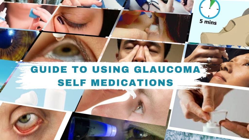
Guide to Using Glaucoma Self Medications image,

A Guide to Self-Administering Glaucoma Eye Drops Effectively

Menu Home About Us Doctor Vision & Mission Services Advanced Cataract Services Cornea Glaucoma Vision Correction/Refractive Surgery Pediatric Ophthalmology Occuloplasty Medical Retina Opticals Conditions We Treat Allergic conjunctivitis Keratoconus Dry Eyes Patient Education Blog Posters Videos Gallery A Guide to Self-Administering Glaucoma Eye Drops Effectively Managing glaucoma involves more than just taking medications it requires mastering the technique of self-administering eye drops. This skill ensures that the medication reaches its intended target and maximizes its therapeutic benefits. Learning how to administer eye drops correctly can significantly impact the progression of glaucoma and help preserve your vision over time. Importance of Proper Eye Drop Administration Efficacy: Proper administration ensures that the medication reaches the eye’s surface and is absorbed effectively to lower intraocular pressure. Minimize Side Effects: Accurate administration reduces the risk of side effects such as eye irritation or systemic absorption of the medication. Vision Preservation: Consistent use of eye drops as prescribed by your eye care provider helps slow down the progression of glaucoma and preserve your vision. Tips and Tricks for Self-Administering Glaucoma Eye Drops Non-Dominant Hand Use your non-dominant hand to gently pull down the lower eyelid. Use your index finger to create a small pocket (lower lid fornix) to catch the eye drop. Dominant Hand Hold the eye drop bottle with your dominant hand. Use your thumb joint as a fulcrum on your index finger to control the squeeze and release of the bottle. Maintaining Distance from Eyelid Keep the eye drop bottle about 1-2 inches away from your eye to prevent accidentally touching the bottle tip to your eye or eyelashes. Practice Aiming Aim the eye drop into the lower lid fornix, not directly onto the cornea or eyelashes. Adjust your aim as you practice to ensure the drop lands in the pocket and not on the eyelashes or cheek. Blinking and Closing Eyes After instilling the eye drop, close your eyes gently. Avoid blinking excessively or squeezing your eyelids tightly, as this can cause the medication to drain away from your eye. Nasolacrimal Duct Technique Immediately after administering the eye drop, press lightly on the inner corner of your eye (near the nose) using your finger. This helps block the tear duct and prevents the medication from draining into your nasal passage and bloodstream. Use of Mirror If you find it challenging to aim the drops accurately, use a mirror to help guide your hand and position. Maintaining Hygiene Wash your hands thoroughly with soap and water before handling your eye drops to prevent introducing bacteria into your eyes. Avoid touching your eyelids or lashes with the eye drop bottle tip to prevent contamination. Recap and Store Properly After using the eye drops, recap the bottle securely to prevent contamination and store it according to the manufacturer’s instructions. Stay Calm and Patient Self-administering eye drops can be tricky at first, but with practice and patience, you’ll become more confident in your technique. Don’t hesitate to ask for help or guidance from your eye care provider if needed. Consistency is Key Stick to a regular schedule for using your eye drops. Set alarms or use medication reminders to ensure you don’t miss doses. Make using your eye drops a part of your daily routine to effectively manage your glaucoma and preserve your vision. Conclusion Mastering the technique of self-administering glaucoma eye drops is crucial for maintaining your eye health and managing your condition effectively. By following these tips and tricks, you can ensure that you administer your medications accurately and minimize the risk of complications. Remember, practice makes perfect—be patient with yourself as you develop your skills. If you have any concerns or questions about your eye drop regimen, don’t hesitate to reach out to your eye care provider for guidance. Together, you can work towards preserving your vision and enjoying optimal eye health. This guide empowers you to take control of your glaucoma treatment by teaching you the essential skills of eye drop administration. With diligence and proper technique, you can effectively manage your condition and protect your vision for years to come. Services & Treatments Cataract Cornea Glaucoma Vision Correction Pediatric Ophthalmology Occuloplasty Medical Retina Opticals Contact info Hanumasai Nagar, Shanti Nagar, Uppal, Hyderabad, Telangana 500039, India +91 80746 61750 shanthinethralaya@gmail.com Opening Hours: 09:00 AM – 08:00 PM Social Profiles Instagram Whatsapp Youtube Icon-map-marker Whatsapp Icon-email Instagram Youtube Icon-map-marker1 Call +91-8074661750

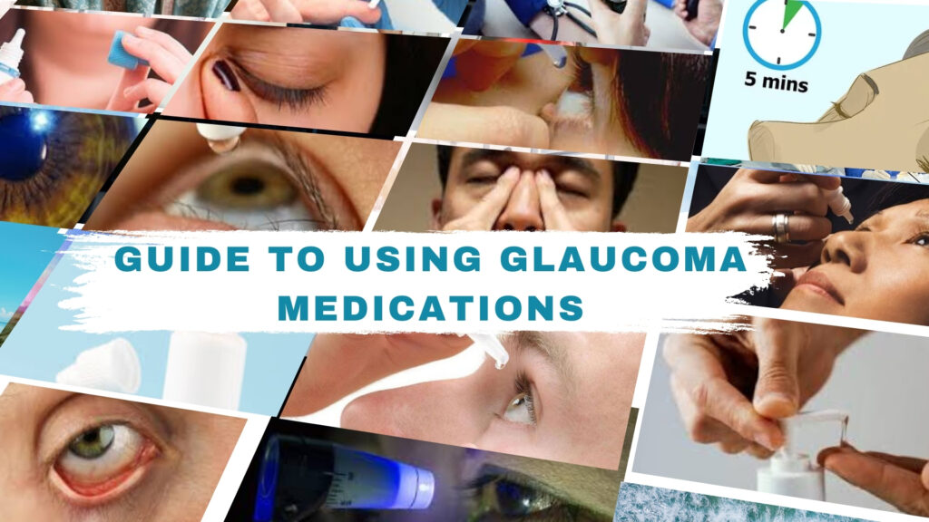
Guide to Using Glaucoma Medications image

Comprehensive Guide to Using Glaucoma Medications or Eye Drops Effectively

Menu Home About Us Doctor Vision & Mission Services Advanced Cataract Services Cornea Glaucoma Vision Correction/Refractive Surgery Pediatric Ophthalmology Occuloplasty Medical Retina Opticals Conditions We Treat Allergic conjunctivitis Keratoconus Dry Eyes Patient Education Blog Posters Videos Gallery Comprehensive Guide to Using Glaucoma Medications or Eye Drops Effectively Managing glaucoma requires consistent use of medications or eye drops to control eye pressure and preserve vision. Proper administration of these medications is crucial for their effectiveness. This guide provides step-by-step instructions on how to self-administer glaucoma eye drops to ensure you get the most benefit from your treatment. Prepare Your Supplies Glaucoma eye drops prescribed by your doctor Tissues or clean cloth Mirror (optional, for better visibility) Hand sanitizer or soap and water for hand hygiene Maintain a Schedule Consistency is key to managing glaucoma effectively. Use your eye drops at the same time(s) each day. Set reminders on your phone or use a medication calendar to track doses and ensure you don’t miss any. Hand Hygiene Wash your hands thoroughly with soap and water or use hand sanitizer to reduce the risk of infection. Dry your hands with a clean towel before handling the eye drops. Position Yourself Correctly Find a stable and comfortable position. Sit down or stand in front of a mirror if needed for better visibility and control. Open the Eye Drop Bottle Twist off the cap carefully to avoid contaminating the tip of the eye drop bottle. Some bottles may require you to break a seal on first use. Tilt Your Head Back or Lie Down Tilt your head back slightly or lie down on a bed or couch with your head supported by pillows. This helps keep the eye drops in your eye and prevents them from running out. Pull Down the Lower Eyelid Using one hand, gently pull down the lower eyelid to create a small pocket. This technique helps prevent the tip of the eye drop bottle from touching your eye, reducing the risk of contamination. Instill the Eye Drop Hold the eye drop bottle upside down close to your eye, but not touching it. Squeeze the bottle gently to release one drop into the pocket formed by your lower eyelid. Avoid blinking or squeezing your eyelids tightly immediately after instilling the drop. Close Your Eyes Close your eyes gently after instilling the drop. This allows the medication to spread evenly over your eye’s surface and reduces drainage into your tear ducts. Apply Pressure to the Inner Corner After closing your eyes, use your finger to apply gentle pressure to the inner corner of your eyelid near your nose (nasolacrimal duct). This helps block the tear duct and prevents the medication from draining into your nasal passage and being absorbed systemically. Use a Tissue or Cloth Use a tissue or clean cloth to gently wipe away any excess medication around your eyes. Avoid rubbing your eyes, as this can cause irritation. Recap the Bottle Replace the cap securely on the eye drop bottle immediately after use to prevent contamination and preserve the medication’s efficacy. Tips for Self-Administering Eye Drops Practice Makes Perfect: It may take a few tries to get comfortable with the process. Practice in front of a mirror if needed. Ask for Help if Needed: If you find it difficult to administer drops on your own, ask a family member or caregiver for assistance. Stay Committed: Consistency is crucial for managing glaucoma effectively. Make using your eye drops a part of your daily routine to ensure optimal treatment outcomes. Conclusion Effectively using glaucoma medications or eye drops is essential for maintaining eye health and preventing vision loss. By following these steps and incorporating proper hygiene practices, you can ensure that your treatment regimen supports your overall eye care goals. If you have any questions or concerns about your medication regimen, don’t hesitate to consult with your eye care provider. Together, you can work towards preserving your vision and enhancing your quality of life. Remember, your eye health is in your hands—literally. Stay diligent and proactive in managing your glaucoma for the best possible outcomes. Services & Treatments Cataract Cornea Glaucoma Vision Correction Pediatric Ophthalmology Occuloplasty Medical Retina Opticals Contact info Hanumasai Nagar, Shanti Nagar, Uppal, Hyderabad, Telangana 500039, India +91 80746 61750 shanthinethralaya@gmail.com Opening Hours: 09:00 AM – 08:00 PM Social Profiles Instagram Whatsapp Youtube Icon-map-marker

Innovations in Glaucoma Treatment explores cutting-edge approaches in glaucoma care.

Innovations in Glaucoma Treatment

Menu Home About Us Doctor Vision & Mission Services Advanced Cataract Services Cornea Glaucoma Vision Correction/Refractive Surgery Pediatric Ophthalmology Occuloplasty Medical Retina Opticals Conditions We Treat Allergic conjunctivitis Keratoconus Dry Eyes Patient Education Blog Posters Videos Gallery Innovations in Glaucoma Treatment Glaucoma, a complex eye disease affecting millions worldwide, poses a significant challenge due to its progressive nature and potential for irreversible vision loss. While managing advanced stages of glaucoma can be daunting, recent innovations in both diagnosis and treatment offer renewed hope and possibilities for enhancing quality of life and preserving vision. Advanced Diagnostics Optical Coherence Tomography (OCT) Optical Coherence Tomography (OCT) has revolutionized glaucoma management by providing detailed, cross-sectional images of the retina and optic nerve. Innovations in OCT technology, such as enhanced depth imaging and swept-source OCT, enable clinicians to detect subtle structural changes earlier. This precision allows for timely intervention to slow disease progression and preserve remaining vision. Artificial Intelligence (AI) in Diagnosis AI-driven algorithms have transformed glaucoma diagnostics by analyzing complex OCT data with unmatched accuracy. These algorithms detect subtle optic nerve and retinal changes indicative of glaucoma, facilitating earlier diagnosis and personalized treatment strategies. By harnessing AI’s capabilities, clinicians can tailor interventions to individual patient needs, offering advanced care even in advanced stages of the disease. Innovative Treatment Modalities Minimally Invasive Glaucoma Surgery (MIGS) Minimally Invasive Glaucoma Surgery (MIGS) represents a significant advancement in surgical options for glaucoma patients, particularly those with advanced disease. These microsurgical procedures, such as micro stent implantation and trabecular bypass devices, aim to improve aqueous humour outflow while minimizing trauma to the eye. MIGS offers potential benefits such as reduced dependence on medications and quicker recovery times compared to traditional surgeries. Types of MIGS Procedures Microstents: Devices placed within the eye’s drainage system to enhance fluid outflow. Trabecular Bypass Devices: Implants designed to bypass the trabecular meshwork, facilitating improved drainage. Suprachoroidal Devices: Devices that enhance fluid drainage through the suprachoroidal space, offering alternative pathways for aqueous humor to exit the eye. Benefits and Considerations MIGS procedures are associated with fewer complications and quicker recovery times, making them suitable options for patients, including those with advanced glaucoma who may be at higher risk for surgery-related complications. Neuroprotection and Regenerative Therapies Research into neuroprotective agents and regenerative treatments aims to preserve optic nerve function and potentially reverse damage in advanced glaucoma. Neurotrophic factors, antioxidants, and gene therapies show promise in protecting retinal ganglion cells (RGCs) and promoting nerve regeneration. These therapies not only aim to halt disease progression but also strive to restore visual function and improve quality of life for patients facing advanced stages of glaucoma. Role of Neuroprotective Agents Neuroprotective agents like brimonidine and memantine have demonstrated potential in reducing oxidative stress and inflammation within the retina, thereby preserving RGC function. These agents complement existing treatments by addressing the underlying mechanisms of neurodegeneration in glaucoma. Advances in Regenerative Therapies Regenerative therapies, including stem cell therapy and gene therapy, hold promise for repairing and replacing damaged retinal cells. These approaches aim to restore visual function by promoting cell survival and regeneration in the optic nerve, offering new hope for patients with advanced glaucoma. Hope and Future Directions Personalized Care: Tailoring Treatment Plans The future of glaucoma care lies in personalized treatment strategies that consider individual patient characteristics, including genetic predispositions and disease progression patterns. By integrating genetic testing and biomarker analysis, clinicians can predict treatment responses more accurately and customize therapies to optimize outcomes while minimizing side effects. Telemedicine: Enhancing Accessibility & Monitoring Advancements in telemedicine and remote monitoring technologies facilitate ongoing care and real-time disease management for glaucoma patients. Virtual consultations, remote IOP monitoring, and mobile health applications enable patients to participate actively in their eye health management from anywhere, ensuring timely adjustments to treatment plans and enhancing overall treatment adherence. Conclusion As we navigate the evolving landscape of glaucoma treatment, these innovations offer tangible hope and possibilities for patients with advanced disease. From advanced diagnostics and minimally invasive surgeries to cutting-edge neuroprotection and regenerative therapies, the field of glaucoma care continues to advance, promising improved outcomes and preserved vision. By staying informed and engaged with their eye care providers, patients can embrace these advancements with confidence, knowing that the future holds promising solutions for managing and potentially reversing the impact of glaucoma. Together, let’s continue to advocate for comprehensive glaucoma care that enhances quality of life and preserves vision for all patients, offering hope and progress in the journey against this challenging disease. Services & Treatments Cataract Cornea Glaucoma Vision Correction Pediatric Ophthalmology Occuloplasty Medical Retina Opticals Contact info Hanumasai Nagar, Shanti Nagar, Uppal, Hyderabad, Telangana 500039, India +91 80746 61750 shanthinethralaya@gmail.com Opening Hours: 09:00 AM – 08:00 PM Social Profiles Instagram Whatsapp Youtube Icon-map-marker

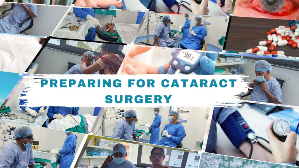
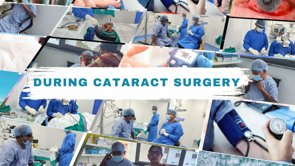
Patients and doctors preparing for cataract surgery at Shanthi Nethralaya Eye Hospital - Pre-surgery medical checks and guidance

Preparing for Cataract Surgery

Menu Home About Us Doctor Vision & Mission Services Advanced Cataract Services Cornea Glaucoma Vision Correction/Refractive Surgery Pediatric Ophthalmology Occuloplasty Medical Retina Opticals Conditions We Treat Allergic conjunctivitis Keratoconus Dry Eyes Patient Education Blog Posters Videos Gallery Preparing for Cataract Surgery As you prepare for your upcoming cataract surgery, it’s important to understand what to expect and how to get ready. This will help ensure everything goes smoothly and that you recover quickly. Let’s go over some key points to help you prepare. Stay Calm Educate Yourself: Understanding the procedure can alleviate much of the anxiety you might be feeling. Cataract surgery is generally quick, minimally invasive, and has a very high success rate. Ask Questions: Feel free to ask me any questions you have. Knowing what to expect can be very reassuring. Practice Relaxation: Simple relaxation techniques, such as deep breathing or meditation, can help you stay calm. Remember, our team will be with you every step of the way. Take All Medications Continue Your Medications: Most patients can continue taking their usual medications right up to the time of surgery. If there are any exceptions, I will let you know. Eye Drops: You might be prescribed an antibiotic or anti-inflammatory eye drops to use before the surgery. Make sure to follow the instructions for these drops carefully. Inform Me About All Medications: Please inform me of all medications, including over-the-counter drugs and supplements, so we can avoid any potential interactions or complications. No Need to Skip Meals Eat a Light Meal: Have a light meal before the surgery. Avoid heavy or greasy foods that might upset your stomach. Stay Hydrated: Drink plenty of water unless you’ve been advised otherwise. Staying hydrated is important for your overall health and well-being. Shave Before Surgery Facial Hair: If you have facial hair, shave it before your surgery. This helps to maintain a clean and sterile environment. Eyebrows and Eyelashes: Leave your eyebrows and eyelashes as they are. We’ll take care of anything that needs to be done in a sterile environment. Other Important Preparations Arrange Transportation: Cataract surgery is an outpatient procedure, meaning you’ll go home the same day. However, you won’t be able to drive yourself home. Arrange for someone to bring you to and from the surgery center. Comfortable Clothing: Wear comfortable, loose-fitting clothing on the day of the surgery. Avoid any accessories like necklaces, earrings, or contact lenses. Remove Makeup and Nail Polish: Ensure your face is free of makeup, lotions, or creams. Remove any nail polish as well, as it can interfere with monitoring devices. Rest Well: Get a good night’s sleep before your surgery. Being well-rested can help you feel more relaxed and better prepared. Plan for Recovery: Set up a comfortable recovery space at home. Stock up on easy-to-prepare meals and ensure you have someone to assist you with daily activities for the first few days. Understand the Procedure Duration: The surgery typically takes about 15-30 minutes. Anesthesia: We use local anesthesia, so you’ll be awake but won’t feel any pain in your eye. Recovery: Most patients experience improved vision within a few days, although complete healing can take a few weeks. Post-Surgery Care Follow-Up Appointments: Make sure to attend all follow-up appointments so we can monitor your healing and address any concerns. Eye Protection: Use the protective shield or glasses we provide to protect your eye from dust, light, and accidental bumps. Avoid Strenuous Activities: Refrain from heavy lifting, bending over, or any activities that could strain your eyes for at least a week. Medication Adherence: Continue using any prescribed eye drops or medications exactly as directed to prevent infection and inflammation. Report Issues: Immediately report any unusual symptoms such as severe pain, vision loss, or excessive redness to us. Conclusion By following these guidelines, you’ll be well-prepared for your cataract surgery. Remember, this procedure is one of the safest and most effective surgeries available today. With proper preparation and care, you can look forward to excellent results and a speedy recovery. If you have any questions or concerns, please don’t hesitate to reach out. We’re here to support you every step of the way. Thank you for entrusting us with your eye care. We look forward to helping you achieve a clearer vision and a better quality of life. Services & Treatments   Cataract Cornea Glaucoma Vision Correction Pediatric Ophthalmology Occuloplasty Medical Retina Opticals Contact info Hanumasai Nagar, Shanti Nagar, Uppal, Hyderabad, Telangana 500039, India +91 80746 61750 shanthinethralaya@gmail.com Opening Hours: 09:00 AM – 08:00 PM Social Profiles Instagram Whatsapp Youtube Icon-map-marker

"Managing Glaucoma at Shanthinethralaya Eye Hospital, Treatment by Dr. B. Shanthi Niketh"

Lifestyle Changes for Managing Glaucoma

Menu Home About Us Doctor Vision & Mission Services Advanced Cataract Services Cornea Glaucoma Vision Correction/Refractive Surgery Pediatric Ophthalmology Occuloplasty Medical Retina Opticals Conditions We Treat Allergic conjunctivitis Keratoconus Dry Eyes Patient Education Blog Posters Videos Gallery Lifestyle Changes for Managing Glaucoma Glaucoma, a progressive eye disease affecting the optic nerve, requires ongoing management to preserve vision and quality of life. Alongside medical treatments, lifestyle modifications play a crucial role in supporting eye health and managing the condition effectively. By adopting healthy habits, individuals with glaucoma can optimize their well-being and potentially slow disease progression. Importance of Lifestyle Changes The choices we make in daily life can impact the progression of glaucoma.Lifestyle modifications aim to reduce intraocular pressure (IOP), enhance circulation to the optic nerve, and promote overall eye health. Integrating these changes into your routine can complement medical treatments and support long-term vision preservation. Key Lifestyle Modifications Diet and Nutrition A balanced diet rich in antioxidants, vitamins (particularly vitamin C and E), and omega-3 fatty acids can benefit eye health. Foods such as leafy greens, citrus fruits, nuts, and fish like salmon or tuna are known to support optic nerve function and reduce inflammation. Limiting caffeine and excessive salt intake may also help manage IOP levels. Exercise and Physical Activity Regular exercise improves blood flow throughout the body, including to the eyes. Engaging in moderate aerobic activities such as walking, cycling, or swimming can help lower IOP and reduce the risk of glaucoma progression. Consult with your healthcare provider to determine safe exercise routines tailored to your condition. Stress Management Chronic stress can elevate IOP levels and potentially accelerate glaucoma progression. Incorporating stress-reducing techniques such as yoga, meditation, deep breathing exercises, or hobbies that promote relaxation can contribute to better eye health. Prioritizing adequate sleep and maintaining a balanced lifestyle are also essential. Joining a Support Community Connecting with others who are managing glaucoma can provide valuable support, shared experiences, and encouragement. Joining a support group or community can offer emotional support, practical tips for coping with the challenges of glaucoma, and opportunities to learn from others’ experiences. Regular Eye Exams: Schedule routine eye check-ups as recommended by your eye care professional to monitor changes in your condition. Medication Adherence: Follow your prescribed treatment regimen diligently, including using eye drops as directed to maintain stable IOP levels. Hydration: Drink plenty of water throughout the day to maintain hydration and support overall eye heTips for Better Eye Care:alth. Quit Smoking: Smoking has been linked to an increased risk of developing glaucoma and can worsen existing conditions. Quitting smoking can improve overall eye health and reduce disease progression risks. Integrating Changes into Your Routine Incorporate these lifestyle modifications gradually into your daily life to ensure sustainability. Keep track of your progress and consult with your healthcare provider regularly to adjust your treatment plan as needed. By prioritizing proactive eye care and adopting healthy habits, you can optimize your vision health and enhance your quality of life with glaucoma. Conclusion Managing glaucoma involves a comprehensive approach that includes medical treatments and lifestyle modifications. By embracing healthy habits such as maintaining a balanced diet, staying physically active, managing stress effectively, and joining a support community, you can support your eye health and potentially slow the progression of glaucoma. Remember, proactive management and regular communication with your healthcare team are key to preserving vision and enjoying a fulfilling life with glaucoma. Embrace these lifestyle changes today to safeguard your vision tomorrow, and empower yourself in the journey of managing glaucoma effectively. Services & Treatments   Cataract Cornea Glaucoma Vision Correction Pediatric Ophthalmology Occuloplasty Medical Retina Opticals Contact info Hanumasai Nagar, Shanti Nagar, Uppal, Hyderabad, Telangana 500039, India +91 80746 61750 shanthinethralaya@gmail.com Opening Hours: 09:00 AM – 08:00 PM Social Profiles Instagram Whatsapp Youtube Icon-map-marker

Surgeon performing cataract surgery at Shanthi Nethralaya Eye Hospital using advanced technology

Exploring Surgical Options for Cataract Surgery

Menu Home About Us Doctor Vision & Mission Services Advanced Cataract Services Cornea Glaucoma Vision Correction/Refractive Surgery Pediatric Ophthalmology Occuloplasty Medical Retina Opticals Conditions We Treat Allergic conjunctivitis Keratoconus Dry Eyes Patient Education Blog Posters Videos Gallery Exploring Surgical Options for Cataract Surgery Best Surgical Options for Cataract Surgery at Shanthi Nethralaya Eye Hospital Cataract surgery is one of the most common surgeries worldwide, helping restore vision by treating the clouding of the eye’s lens. With advancements in techniques and technology, patients now have more options, allowing for customized treatments to fit their specific needs. This guide covers the different surgical options, from traditional to advanced methods, and offers tips on choosing the right surgeon and healthcare provider Phacoemulsification Surgery Phacoemulsification, commonly known as phaco, represents the gold standard in cataract surgery. This technique involves creating a small incision in the cornea through which an ultrasonic device is inserted to fragment and remove the clouded lens. Phacoemulsification offers numerous benefits, including minimal incision size, faster visual recovery, and precise lens removal, making it the preferred choice for many patients and surgeons alike. Manual Small-Incision Cataract Surgery (MSICS) Manual small-incision cataract surgery (MSICS) serves as an alternative to phacoemulsification, particularly in settings with limited access to advanced technology. In MSICS, a larger incision is made to facilitate the removal of the cataract in one piece, making it suitable for cases with dense or mature cataracts. While less common in developed countries, MSICS remains a vital option in regions where resources are constrained. Femtosecond Laser-Assisted Cataract Surgery (FLACS) Femtosecond laser-assisted cataract surgery (FLACS) combines the precision of laser technology with the benefits of phacoemulsification. The laser is used to create precise incisions in the cornea, fragment the cataract, and soften the lens for easier removal. FLACS offers enhanced accuracy, customization of incision parameters, and reduced energy requirements, potentially leading to improved visual outcomes and faster recovery for patients. Refractive Cataract Surgery Refractive cataract surgery goes beyond simply removing the cataract by addressing pre-existing refractive errors, such as nearsightedness, farsightedness, and astigmatism. Techniques in refractive cataract surgery include implantation of astigmatism-correcting toric intraocular lenses (IOLs), multifocal or extended depth of focus (EDOF) IOLs, and accommodating IOLs, providing patients with improved vision at multiple distances and reduced dependence on corrective lenses. Considerations and Precautions Several factors must be considered when undergoing cataract surgery, including patient suitability, surgical expertise, and post-operative care. Patients should undergo thorough evaluations to determine the most appropriate surgical approach, choose experienced surgeons and reputable healthcare providers, and adhere to prescribed medication regimens and follow-up appointments to ensure a smooth recovery process. Why Choose Shanthi Nethralaya Eye Hospital and Dr. B. Shanthi Niketh for Your Cataract Surgery Expertise and Experience Dr. B. Shanthi Niketh is a distinguished ophthalmologist with extensive experience in cataract surgery and other advanced ophthalmic procedures. The hospital’s team comprises highly skilled professionals dedicated to providing the highest standard of care. State-of-the-Art Facilities Shanthi Nethralaya Eye Hospital boasts cutting-edge technology and modern infrastructure to facilitate advanced cataract surgery techniques, ensuring optimal outcomes and patient safety. Personalized Care and Attention Patients at Shanthi Nethralaya receive individualized care tailored to their specific needs and preferences. Dr. B. Shanthi Niketh and his team prioritize thorough evaluations, open communication, and patient comfort throughout the treatment process. Comprehensive Services In addition to cataract surgery, Shanthi Nethralaya Eye Hospital offers a wide range of ophthalmic services, including comprehensive eye examinations, treatment for various eye conditions, and refractive surgery options, providing holistic care under one roof. Positive Patient Feedback and Reputation Shanthi Nethralaya Eye Hospital has garnered a reputation for excellence in eye care, supported by numerous positive patient testimonials and reviews. Dr. B. Shanthi Niketh’s compassionate approach and commitment to patient satisfaction make him a trusted choice for cataract surgery. In conclusion, choosing the right surgical option for cataract surgery requires careful consideration of various factors, including patient needs, surgical techniques, and healthcare providers. Whether opting for traditional phacoemulsification, advanced FLACS, or refractive cataract surgery, patients can trust in the expertise, state-of-the-art facilities, and personalized care offered by Shanthi Nethralaya Eye Hospital and Dr. B. Shanthi Niketh to achieve optimal visual outcomes and regain clarity in their eyes. Services & Treatments Cataract Cornea Glaucoma Vision Correction Pediatric Ophthalmology Occuloplasty Medical Retina Opticals Contact info Hanumasai Nagar, Shanti Nagar, Uppal, Hyderabad, Telangana 500039, India +91 80746 61750 shanthinethralaya@gmail.com Opening Hours: 09:00 AM – 08:00 PM Social Profiles Instagram Whatsapp Youtube Icon-map-marker

"Cataract surgery at Shanthinethralaya Eye Hospital, performed by Dr. B. Shanthi Niketh"

Choosing the Right Intraocular Lens (IOL) for Cataract Surgery

Menu Home About Us Doctor Vision & Mission Services Advanced Cataract Services Cornea Glaucoma Vision Correction/Refractive Surgery Pediatric Ophthalmology Occuloplasty Medical Retina Opticals Conditions We Treat Allergic conjunctivitis Keratoconus Dry Eyes Patient Education Blog Posters Videos Gallery Choosing the Right Intraocular Lens (IOL) for Cataract Surgery Cataract surgery is one of the most common and successful procedures performed today. During the surgery, the cloudy natural lens of your eye is replaced with a clear artificial lens called an intraocular lens (IOL). This can significantly improve your or your loved one’s vision quality and overall quality of life. Importance of Choosing the Right IOL Choosing the right IOL is crucial because it will affect your vision after surgery. With various types of IOLs available, understanding their differences and how they can meet your needs is essential. Understanding Intraocular Lenses (IOLs) What is an IOL? An intraocular lens (IOL) is an artificial lens placed inside your eye to replace the natural lens removed during cataract surgery. Modern IOLs are designed to last a lifetime and are made from materials such as acrylic. Types of IOLs Based on Functionality Monofocal IOLs These lenses have a single focal point, usually set for clear distance vision. You might still need glasses for near or intermediate tasks. Multifocal IOLs These lenses provide multiple focal points, allowing you to see clearly at near, intermediate, and far distances, potentially reducing your need for glasses. Aspheric IOLs These lenses are designed to reduce spherical aberrations, providing sharper vision, especially in low-light conditions. Hydrophilic Acrylic IOLs Material: Hydrophilic acrylic, known for its flexibility and ease of implantation. Advantages: High water content makes them more flexible, easier to handle, and provides better biocompatibility. Disadvantages: Higher risk of posterior capsule opacification compared to hydrophobic IOLs. Hydrophobic Acrylic IOLs Material: Hydrophobic acrylic, which repels water and proteins. Advantages: Lower incidence of posterior capsule opacification, better resistance to glistening (small water inclusions). Disadvantages: Can be less flexible, slightly more prone to glistening than hydrophilic IOLs. Extended Depth of Focus (EDOF) IOLs These lenses offer a continuous range of vision from distance to intermediate, with some degree of near vision, aiming to reduce visual disturbances like halos and glare. Toric IOLs Designed specifically to correct astigmatism, these lenses can provide you with sharper vision if you have significant corneal astigmatism. Types of IOLs Based on Material Polymethylmethacrylate (PMMA) IOLs Material: PMMA, a rigid plastic. Advantages: Excellent optical clarity and biocompatibility. Disadvantages: Requires a larger incision for implantation due to its rigidity. Types of IOLs Based on Surface Design Aspheric IOLs: These lenses are designed to reduce spherical aberrations, providing sharper vision, especially in low-light conditions. Advantages: Improved contrast sensitivity and better overall visual quality. Disadvantages: Can be more expensive than traditional spherical IOLs. Types of IOLs Basedand on Hydrophilicity Hydrophilic Acrylic IOLs Material: Hydrophilic acrylic, known for its flexibility and ease of implantation. Advantages: High water content makes them more flexible, easier to handle, and provides better biocompatibility. Disadvantages: Higher risk of posterior capsule opacification compared to hydrophobic IOLs. Hydrophobic Acrylic IOLs Material: Hydrophobic acrylic, which repels water and proteins. Advantages: Lower incidence of posterior capsule opacification, better resistance to glistening (small water inclusions). Disadvantages: Can be less flexible, slightly more prone to glistening than hydrophilic IOLs. Factors to Consider When Choosing an IOL Profession or Visual Needs Night Driving Near Vision Needs Computer Work   Night Driving Near Vision Needs Computer Work Presence of Astigmatism If you have significant astigmatism, toric IOLs can correct this refractive error, providing you with clearer vision without the need for additional glasses for astigmatism. Tolerance for Visual Disturbances Multifocal and some EDOF IOLs can cause visual disturbances such as halos and glare, especially at night. If you are sensitive to these issues, you might prefer monofocal or toric IOLs. Pre-existing Eye Conditions Corneal Scars Scarring on your cornea can affect the clarity of your vision and may influence the type of IOL best suited for you. High Myopia If you are highly nearsighted, specific IOLs may be better suited to correct your vision effectively. Retina Problems Including Macular Degeneration These conditions can affect how well you see with certain types of IOLs. It’s important to choose an IOL that complements the health of your retina. Diabetic Eye Disease Diabetes can lead to various eye problems that might affect IOL selection, weighing factors like visual acuity, contrast sensitivity, and potential impact on diabetic retinopathy progression. Optic Nerve Issues Glaucoma and other optic nerve problems can influence IOL selection, as these conditions can affect overall visual quality. Your Postoperative Goals and Expectations It’s important to discuss your goals and expectations for vision after surgery. Some people are comfortable with occasionally using glasses, while others seek the most glasses-free vision possible. Consultation with an Ophthalmologist Your Visual Needs We will discuss your daily activities and visual preferences to help tailor the IOL selection to your lifestyle. Eye Measurements Biometry: This measures the length of your eye and the curvature of your cornea. Accurate biometry is crucial for determining the appropriate IOL power. Corneal Topography: This maps the surface curvature of your cornea, helping to identify any irregularities or astigmatism that need correction with a toric IOL. Keratometry: This measures the curvature of the front surface of your eye, further aiding in calculating the correct IOL power. Ocular Coherence Tomography (OCT): This imaging test provides detailed images of your retina and macula, ensuring they are healthy and capable of supporting a specific type of IOL. Endothelial Cell Count: This counts the cells on the inner layer of your cornea, which can affect your eye’s ability to tolerate surgery and certain IOLs. Medical History We will review any existing eye conditions, general health issues, and medications that might impact your eye health or surgery outcomes. Conclusion Summary of Key Points Choosing the right IOL involves considering your vision needs, lifestyle, astigmatism, tolerance for visual disturbances, and eye health. Decision-Making A thorough consultation with an ophthalmologist is essential to make an informed decision

"Various stages of cataract surgery, including patient preparation, surgery, and post-operative care, with doctors and nurses assisting, and the text 'During Cataract Surgery' displayed.

A Day in the Life: What Happens During Cataract Surgery

Menu Home About Us Doctor Vision & Mission Services Advanced Cataract Services Cornea Glaucoma Vision Correction/Refractive Surgery Pediatric Ophthalmology Occuloplasty Medical Retina Opticals Conditions We Treat Allergic conjunctivitis Keratoconus Dry Eyes Patient Education Blog Posters Videos Gallery A Day in the Life: What Happens During Cataract Surgery Hello! If you’re preparing for cataract surgery, it’s natural to feel a mix of anticipation and a bit of nervousness. I’m here to walk you through what happens on the day of your surgery, from pre-op to the procedure itself, and finally, to your post-op recovery. Understanding each step can help you feel more at ease. Pre-Operative Preparation Arriving at the Hospital or Surgical Center Check-In: When you arrive, you’ll check in at the reception. Please arrive on time as indicated in your appointment details. Paperwork: We may need you to complete some final paperwork, including consent forms. Make sure to bring your ID, insurance details, and any relevant medical documents. Pre-Op Room Changing Clothes: We’ll provide you with a hospital gown to change into. Wear comfortable clothing and leave valuables at home. Vital Signs Check: A nurse will check your blood pressure, pulse, and temperature to ensure you’re ready for surgery. Eye Drops: You’ll receive special eye drops to dilate your pupils and numb your eyes. This topical anesthesia ensures you won’t feel pain during the procedure. Meeting the Surgical Team Consultation: We’ll have a brief consultation to review the procedure and address any last-minute questions you may have. Marking the Eye: I’ll mark the eye that we’ll be operating on to avoid any confusion. The Surgery Arriving at the Hospital or Surgical Center Preparation: You’ll be taken to the operating room and asked to lie down on a comfortable surgical bed. Your head will be positioned properly, and a sterile drape will be placed around your eye. Reassurance: I’ll be talking to you throughout the procedure to ensure you’re comfortable and at ease. During the Surgery Entering the Operating Room:You’ll be guided into the operating room and asked to lie down comfortably. Stay Calm and Focused:During the surgery, keep your focus on the light above you. Stay relaxed and remember, I’ll be here throughout to ensure your comfort. Communication is Key:If you need anything or feel uncomfortable at any point, just let me know. I’m here to assist and ensure your experience is as smooth as possible. Duration The entire surgery usually takes about 15-30 minutes per eye. If you need surgery on both eyes, they will be scheduled on different days. Post-Operative Recovery Immediate Post-Op Care Eye Shield: An eye shield or patch will be placed over your eye to protect it from dust, light, and accidental rubbing. Post-Op Instructions: You’ll receive detailed instructions on how to care for your eye, including the use of prescribed eye drops to prevent infection and reduce inflammation. Going Home Transportation: Ensure you have someone to drive you home, as you won’t be able to drive yourself immediately after the surgery. Rest at Home: Plan to rest for the remainder of the day. Avoid any strenuous activities and try to keep your head elevated. At-Home Care and Follow-Up First 24 Hours Eye Drops: Use the prescribed eye drops as instructed. These drops help with healing and prevent infection. Avoid Touching: Do not touch or rub your eye. If your eye feels itchy or uncomfortable, consult your doctor before taking any action. Follow-Up Appointments Next-Day Check-Up: You’ll have a follow-up appointment the day after your surgery for me to examine your eye and ensure it’s healing properly. Additional Check-Ups: Further follow-up visits will be scheduled to monitor your recovery and vision improvement. Long-Term Recovery Gradual Improvement: Your vision may be blurry initially, but it should start to improve within a few days. Full recovery and optimal vision might take several weeks. Activities: Avoid activities that could strain your eye, such as heavy lifting, bending over, or vigorous exercise. Follow my advice on when you can resume normal activities. Possible Side Effects Common Symptoms: Mild discomfort, itching, and light sensitivity are normal and should subside within a few days. When to Call the Doctor: If you experience severe pain, vision loss, flashes of light, or an increase in redness, contact me immediately. Conclusion Cataract surgery is a straightforward and effective procedure with a high success rate. By understanding what happens on the day of surgery and following the recommended post-op care, you can ensure a smooth recovery and greatly improve your vision. Remember to follow my advice, attend all follow-up appointments, and take care of your eye to enjoy the best possible outcome. If you have any questions or concerns, don’t hesitate to reach out to me. Services & Treatments Cataract Cornea Glaucoma Vision Correction Pediatric Ophthalmology Occuloplasty Medical Retina Opticals Contact info Hanumasai Nagar, Shanti Nagar, Uppal, Hyderabad, Telangana 500039, India +91 80746 61750 shanthinethralaya@gmail.com Opening Hours: 09:00 AM – 08:00 PM Social Profiles Instagram Whatsapp Youtube Icon-map-marker

Scroll to Top